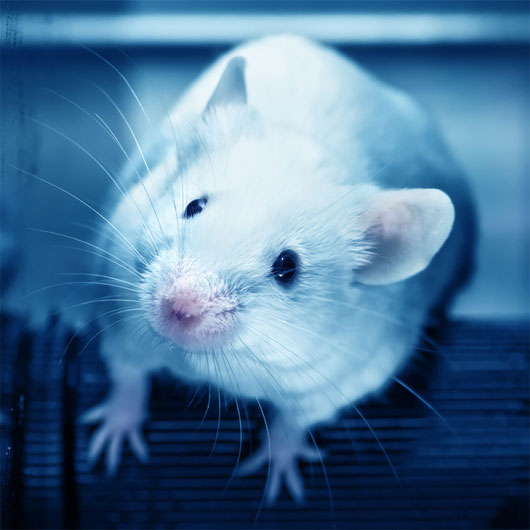
Stress hormones alter brain tissue in mice
(Johns Hopkins researchers) say they have confirmed the suspicions that DNA changes in the blood of mice are exposed to high levels of stress hormones - and denote credits. The effect of anxiety - is directly related to changes in their brain tissue.
This study was presented online before the June print edition in Psychoneuroendocrinology magazine, which introduced what the research team called: the first evidence that epigenetic changes alter the way functions are The gene without changing its basic DNA sequence - and detected in the blood - reflects changes in brain tissue associated with mental illnesses.
This new study only reports epigenetic changes with a single stress response gene called FKBP5 , linked to depression, bipolar disorder and post-traumatic stress disorder. But scientists say they have found the same brain and blood in dozens of genes, which regulate many important processes in the brain.
'Many human studies are based on the assumption that epigenetic changes that occur in the brain - mostly inaccessible and difficult to test - also occur in the blood, possibly Easy check , ' said lead researcher Richard S. Lee, Ph.D., lecturer at the Department of Psychiatry and Behavioral Science at Johns Hopkins University School of Medicine. 'Research done on this mouse shows that blood can tell us what is happening in the brain logically, which is what we had previously assumed, and can help us. better detection and better treatment of mental disorders, as well as providing a better experimental way to check the effectiveness of the medications that are being used ".

In this study, the Johns Hopkins team studied mice with Cushing's disease, a disease caused by excessive production and release of cortisol, a basic stress hormone, also called glucocorticoid . Within 4 weeks, the mice received different doses of stress hormones in their drinking water to evaluate epigenetic changes to the FKBP5 gene.
The researchers took blood samples from mice weekly to assess changes and then examined the rat's brain at the end of the month to see what changes were occurring in the hippocampus brain area due to glucocorticoid exposure. In both mice and humans, the hippocampus brain region plays an important role in memory formation, the ability to store and organize information.
Measurements and analyzes showed that rats received more stress hormones, greater epigenetic changes in the blood and brain tissue, although scientists said changes in the brain occurred in a different part of the gene. with anticipation. This makes finding the connection between blood and brain very difficult, Lee said.
In addition, the more hormones cause stress, the more RNA from the FKBP5 gene is expressed in the blood and brain, and the greater the association with depression. However, potential epigenetic changes are proven to be stronger. This is important, because while RNA levels can be normal after stress hormone levels decrease or change due to minor fluctuations in hormone levels, epigenetic changes are durable (persistent). , reflect overall stress hormone exposure and predict the amount of RNA that will be produced when the level of stress hormone increases.
The team used a previously developed epigenetic assay in their laboratory, requiring only one drop of blood to evaluate the overall stress hormone exposure over 30 days. High levels of stress hormone exposure are considered a risk factor for mental illness in humans and other mammals.
Other Johns Hopkins researchers involved in the study include: Erin R. Ewald; Gary S. Wand: medical doctor; Fayaz Seifuddin, MS Xiaoju Yang: medical doctor; Kellie L. Tamashiro: PhD; and Peter Zandi, Dr. James B. Potash, MD, MPH, a former researcher at Johns Hopkins, also contributed to this study.
The study is funded by grants from National Institutes of Health's National Institute on Alcohol Abuse and Alcoholism (UO1 AA020890) and many other organizations.
- Mini-rat brains in mice raise concerns about intelligent hybridization
- Stress can make your brain smaller
- A scary discovery in genetics
- Successfully developed three-dimensional structural tissue
- Depressed mother gave birth to many stress hormones
- What happens to your brain and body when you're angry?
- Eating candy helps reduce stress
- The mole rats eat the mouse rat manure to get instructions for raising children
- Why are women always beautiful in the eyes of a lover?
- Detecting brain response mechanisms with stress
- Successfully cultivate human intestinal tissue on mice
- Create anti-allergic immune cells from rat fat tissue
 Green tea cleans teeth better than mouthwash?
 Death kiss: This is why you should not let anyone kiss your baby's lips
 What is salmonellosis?
 Caution should be exercised when using aloe vera through eating and drinking