Testicular cancer: Causes, symptoms and treatment
Testicular cancer is a rare cancer in men, accounting for about 1% of all cancers in men.
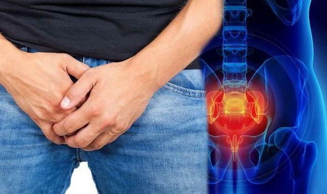
What is testicular cancer?
Testicular cancer occurs when healthy cells in the testes are changed. Healthy cells grow and divide in an orderly manner to keep the body functioning normally. However, sometimes there are abnormal growth cells that make this growth out of control - cancer cells continue to divide into a testicle tumor, which the patient usually feels clinically on. .
Is testicular cancer dangerous ? The good news is that testicular cancer is highly curable if detected and intervened early. For all stages, it is possible to cure 90% of patients. The 5-year survival rate in testicular cancer patients is greater than 95%.
Currently the cause of testicular cancer has not been clearly identified.
Causes of testicular cancer
Currently the cause of testicular cancer has not been clearly identified. Several studies show that the disease stems from the abnormal growth and division of cells in the testes. They grow rapidly uncontrollably, then gradually become tumors in the testes.
More than 90% of testicular cancer begins with stem cells (cells that produce immature sperm) and the cause of their abnormal growth is unknown.
Symptoms of testicular cancer
The most common sign is that the patient finds the scrotum enlarged or palpates in the scrotum. The tumor may be painful or painless.
In addition, the patient has the following symptoms:
- Dull ache in the groin area or lower abdomen
- Scrotum feeling heavy, congestion causes discomfort or pain
- Backache
- May have inguinal lymphadenopathy
Risk factors for testicular cancer include:
- People with hidden testes: the risk of infection is 2.5 to 11 times higher than that of the normal person.
- People with abnormal testicle development (testicular atrophy or undeveloped
- Have family members with testicular cancer
- Race: whites are at higher risk for testicular cancer.
- Several other factors: a history of mumps, testicular hydroceles, inguinal hernia.
- The best way to prevent and detect testicular cancer early is to test yourself.
- Check your testicles once a month, most simply after each shower.
- Test the testicles with two hands, the middle finger on the bottom and the thumb on the testicle. Leveling gently on both sides of the testes, checking the epididymis. The most common location for testicular tumors is on the sides.
- No smoking, no drinking alcohol
- Eat and practice science
- Periodic health examination: healthy people should be examined every 6 months. People with risk factors should have a follow-up visit more often.
Diagnosis of testicular cancer is based on clinical and subclinical signs:
- Scrotum ultrasound: Detects 75% of tumors, determine whether or not hydrocele.
- Ultrasound or abdominal computed tomography: reveals hidden testes, metastatic abdominal lymph nodes.
- Histopathology: diagnostic diagnosis, can be performed immediately after surgery or immediate biopsy during surgery.
- Chest X-ray: helps detect lung metastases
- Tumor markers: AFP, HCG and LDH.
- Bone scans if bone damage is suspected.
Treatment of testicular cancer
Treatment for testicular cancer depends on the type of cancer (testicular tumor, non-testicular tumor) and the stage of the disease. Treatment includes methods: surgery, radiation and chemotherapy.
U planet
- This type of tumor is sensitive to radiation therapy.
- In the early stage, the patient is radiotherapy into the area below the diaphragm, mainly into the inguinal pelvic lymph node and the aorta
- Later stages can use auxiliary chemicals.
U is not seminal
- Surgery with lymphadenectomy is used in cases of small tumors and non-sperm cancer.
- Radiation therapy if the tumor has spread to the adjacent lymph nodes
- Chemical if the disease has spread far.
Follow up after treatment
- Clinical examination, pelvic and abdominal CT scan every 3 months in the first year, then every 6 months for the next 2 years and then once a year.
- Chest X-ray and chest CT if clinically suspected.
- 96% of cases of testicular cancer have been cured
- The cancer has no symptoms early
- Higher men are more likely to get testicular cancer
- New findings related to testicular cancer, heart disease
- Early detection of male cancer should not be overlooked
- Dangerous cancer - cause and prevention
- What is metastatic cancer?
- Biliary cancer - the cause, symptoms and treatment
- The bizarre device detects the most malignant form of cancer for men
- Retinoblastoma: Causes, symptoms, diagnosis and treatment
- Stages of tongue cancer and treatment
- The number of 'breeds' of men is decreasing
 New drug causes cancer to 'starve'
 Why is Australia the country with the highest cancer rate in the world while Vietnam ranks 100th?
 Common cancers in men
 America's incredible discovery: The most feared cancer cell is love