Who invented the layered computer tomography?
Today computers have become very popular in every social activity. But more than 30 years ago, the successful study of computerized layered tomography (VTCLPT) opened a revolution in the field of medical imaging diagnosis, contributing greatly to early detection to treat many incurable diseases.

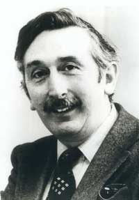
GS.Godfrey N. Hounsfield
With achievements on VTCLPT, two British and American scientists were awarded the Nobel Prize in Medicine in 1979.
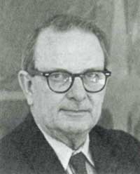
Prize winner: GS. ALLAN M. CORMACK & GS. GODFREY NEWBOLD HOUNSFIELD
The 1979 Nobel Prize for Medicine was awarded to GS. Allan M. Cormack and Godfrey N. Hounsfield for their contributions to the development of computerized tomography, revolutionizing radiotherapy as well as diagnosing neurological diseases.
Allan M. Cormack is a professor of physics at Tufts University (Massachusetts-USA). He was the first to analyze the conditions to make it clear how the right tomography was taken in the biological system and published in 1963-1964, contributing to the development of the theory of computer tomography based on the foundation. on the use of X-rays.
Godfrey Hounsfield is the director of Middlesex's Electrical and Music Industry Research Division (UK), who performed the first computer tomography computer used in medicine in 1968. The patent was granted in 1972. Hounsfield's system helps to diagnose brain and brain imaging, creating a premise to help develop more computerized tomography systems later with technical innovations to help analyze images faster.
Research works

GS.Allan M. Cormack
X rays when passing through organs will give images on film. The image depends on the structure of the tissues of the body that X-rays pass through and are often unclear in depth, so additional shots are needed either face to face. Moreover, film reading also depends on the professional capacity of the photographer and the abnormal nature of the pathology. So the inevitable X-ray results are subjective and misleading. Therefore, the technique of stratified tomography (tomography, in Greek tomos = cut, graph = recorded) has been further developed to isolate the diseased organ image. Although there is progress, it is still not satisfied with the need for early diagnosis by images.
In addition, X-rays have limitations such as being unable to use X-rays more than 25% and X-ray films lack the necessary sensitivity corresponding to tissue thickness. Since then, the idea of using computers to support tomography is formed to solve the problem. And in just 6 years, it has become a revolutionary event in the field of medical imaging diagnostics. By multidimensional scanning through the cross-section of the body to be diagnosed, the image will be recorded by a magnetic resonance induction transducer and then analyzed by electronic calculator. The pre-programmed computer is intended to quickly reproduce the clear, partial image of the matrix-based survey agency on the screen. The reconstructed images will not affect other images, not obscuring each other to avoid confusion. Therefore, computerized magnetic resonance tomography can detect small changes in the tissue structure, while increasing the accuracy by hundreds of times compared to X-rays.
The value of research work
The first tomography computer was built to diagnose neurological diseases of the brain thanks to its sensitivity and accuracy. Since then it is easy to detect neuropathy and changes in brain structure. The location and size of the changes as well as the nature of the vulnerability can be determined, drawing suspicious points to enlarge and display on the screen. Thus determining the nature of many diseases such as cerebral hemorrhage, cerebral vascular accident, cerebral anemia, tumors, encephalitis, cerebral palsy, brain defects . The invention is meaningless As the basis for developing further methods for brain tumors surgery, the details are recorded in memory during brain surgery.

Magnetic resonance imaging technique
This technique does not cause any discomfort to the patient during the scan, and can also refer to computer-based diagnostic diagnostic centers to coordinate diagnosis, treatment, and study of neurological pathologies. The development of digital diagnostics will be complemented by later higher techniques such as ultrasound, radioisotope diagnosis with gamma ray cameras. The most important area of application is the development of radiation therapy for brain tumors.
So far, the most difficult thing about radiation therapy is to clearly identify the location, size and formation and progress of the tumor in the deepest areas of the brain or body, distinguishing tumors with surrounding tissues. With computerized tomography, the therapist will easily recognize where radiation is needed with the optimal beam quality. When the tumor shrinks in treatment will also be clearly recognized on the computer screen, the direction of the beam can change position so that the tumor receives the beam much and does not damage the surrounding tissue. Thus computerized tomography has opened up a new area for radiotherapy to treat cancer.
Applied in current medical imaging diagnosis
From the research work of the 60s and awarded the Nobel Prize in Medicine in the 70s, the medical diagnostic imaging industry has made great progress thanks to the support of informatics.

Compact computer X-ray machine
Hologram of body activities provided by the spiral scanner method will be stored in CD-Rom to detect abnormal structures in the body. New technique for diagnosing breast cancer thanks to digital ultrasound, showing true color images showing blood flow through tissues, supporting the diagnosis of early detection of breast cancer without the need for biopsy to check determined.
Magnetic resonance imaging (Magnetic Resonnance Imaging: MRI) is increasingly developing with information stored in computers, enabling reconstruction of internal organs with high accuracy and high contrast. . Thus the internal body activities are clearly seen as the beating of the heart, tumors in the ovaries, uterus . Blinking techniques with radioactive substances help provide vivid images of moving bodies, as well as digital ultrasound machines, help radiologists to distinguish the shape and structure of the tumor with a true color that shows blood flow through the tissue .
In Vietnam, the computer X-ray machine is compact (CR-Computer Radiology) with the time from shooting to having the image shortened to 70 seconds, taking on a phosphorus plate with a beam dose of only 50% before. The image taken by Scan plate phosphor and processed into the computer has also been applied.
With these benefits, the 1979 Nobel Prize in Medicine awarded the work of using imaging diagnostic computers as a revolutionary breakthrough in the treatment of incurable diseases.
- Super computer scanners
- Siemens introduces a breakthrough in the field of tomography
- Russian scientists created a scanner that could detect even the smallest crack in an airplane
- Denmark invented self-rehabilitation computer
- Software supports tomography to detect lung cancer
- Hitachi developed mobile brain tomography equipment
- Scientists freeze two grasshoppers that are 'forming' to study
- Researching human ancestors with a tomography machine
- Brain scans may soon detect suicidal intent
- The person who invented the email just died at the age of 74
- Toshiba 1TB SSD drives are as small as stamps
- Determine the power of love thanks to ... tomography
 Daily use inventions come from universities
 Special weight loss device helps prevent appetite
 8 inventors were killed by their own inventions
 Iran invented a motor car powered by water