Intrusion strategy of the world's largest virus
The study, conducted by the Weizmann Institute, provides important insights into the process of virus infection. Published online in PLoS Biology, the study revealed amoeba cell invasion mechanisms of Mimiviruses ( 'imitating' viruses) - a name derived from the idea that Mimiviruses mimic bacteria in many ways. dynamic.
The process of living virus-infected cells goes through 2 steps. First, the virus enters the cell. Then, in the second key step, the cells produce new viruses that quickly spread and invade other cells. In the early stages of virus production, the cell forms the outer shell for the virus with a number of proteins, so the shell also has the name of the protein shell. Then the cell copies the viral DNA and then puts it into the shell. The result is a fully functional new virus, ready to leave host cells to attack other cells.
Understanding how to infect viruses and produce viruses during infection helps scientists stop this cycle, limiting viral diseases. However, one of the main problems is the intrusion strategy of different viruses always changing differently.
The Mimivirus is different from other viruses in its special size, which is 5 to 10 times larger than the normal virus thus creating an interesting obstacle in this study. Mimivirus was only discovered in the late 20th century, its unusual size made it impossible to detect by conventional methods. In addition, it contains more genetic material than other viruses - a trait that makes the mimivirus develop effective methods to inject its DNA into host cells as well as introduce newly created genetic material. protein shell during virus production in host cells.

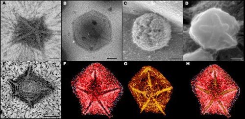
(Fig. A) Conductive electron micrograph (TEM) of the extracellular part of the Mimivirus is fixed, stained exposing the stellar structure.(Fig. B) Cold conduction electron microscopy (Cryo-TEM) Immature Mimivirus is glazed.(Figure C) Extracellular electron microscopy (SEM) microscopic structure of an adult Mimivirus.(Figure D) Cold electron scans (Cryo-SEM) non-immature grainy virus.(Fig. E) Radial slice of the intracellular part of Mimivirus 12 hours after infection.At this stage, parts of the virus flood the host cell.(Fig. F and G) After the virus recovers the volume in Figure E, the polyhedron-shaped shell exposes the extracellular part (red) and the intracellular part (orange).Star structures are present in both types of shells but accept a star-shaped structure enclosed in both extracellular and intracellular in a predetermined order.(Figure H) The two shells (F) and (G) overlap.(Photo: Distinct DNA Exit and Packaging Portals in the Virus Acanthamoeba polyphaga mimivirus Zauberman N, Mutsafi Y, Halevy DB, Shimoni E, Klein E, et al. PLoS Biology Vol. 6, No. 5, e114 doi: 10.1371 / journal. pbio.0060114)
Weizmann Institute Professor - Abraham Minsky - together with graduate student Nathan Zauberman and Yael Mutsafi (Organic Chemistry) collaborated with Dr. Eugenia Klein, Eyal Shimoni (Department of Chemical Research Support) to find a detail. Number of methods used by the virus.
In their new study, scientists for the first time recorded the first three-dimensional image when the genetic material of the virus was introduced into infected cells as well as the image of the process of introducing genetic material. into protein shell. All previous virus studies, scientists believe that the genetic material of the virus enters the cell (during infection) as well as entering the newly formed protein shell (through the production process). new virus inside the cell) through the same conduction channel created in the virus envelope. In contrast, the new study discovered that giant mimiviruses use different conduction channels located in different places on the shell to accomplish these two purposes. They also found that the DNA helix in both processes does not form long chains like other viruses but is separated into dense concentration blocks. They believe that these unique characteristics effectively support both the process of host cell infection as well as the transfer of large amounts of genetic material into the mimivirus shell.
In the study, the electron microscope image recorded the scene of an amoeba invasion of mimivirus showing that immediately after invasion, the walls of the polygonal protein shell consisted of 20 reassembled triangles separated like petals to form an invasive structure of large stars with the nickname 'stargate'. The underlying stargate membrane merges with the amoeba cell membrane, forming a large conduction channel that sends the virus inside the cell. The pressure created by this "abrupt breakdown" is 20 times greater than the pressure when we open the champagne stopper, pushing the viral DNA through the transmission channel. Large sizes allow the genetic material of the virus to move quickly into the amoeba cell.
Other images show how the genetic material of the virus is transferred into the protein shell when the virus first forms in the host cell. In this process, the genetic material of the virus moves to the appropriate location through a gap in the new shell facing the stargate. The transfer of genetic material must overcome the pressure inside the shell. Perhaps it is controlled by an 'engine' inside the shell that hides the entrance.
Scientists believe that the study of the life cycle of mimiviruses, from the infective stage to the production of new viruses, will provide valuable information about the mechanism of action of many other viruses, including viruses that cause illness in humans.
- The biggest virus in the world is exposed
- The virus of blue ear pig disease changes quickly
- Check out the most dangerous viruses on the planet
- Saline intrusion deep in the dry season of 2013
- Southeast Asia should learn Chinese technology strategy
- The most serious disaster in 100 years is raging in the West
- The truths always amaze you about the biological world
- Effective against saline water by manual methods
- HIV virus is no longer as dangerous as before
- France blocks 'gate connecting the worlds'
- Discover the world's most
- Biofuels: An appropriate strategy?
 Why do potatoes have eyes?
 'Tragedy' the world's largest carnivorous life: Death becomes ... public toilet
 Tomatoes were once considered 'poisonous' for 200 years
 Detecting microscopic parasites on human face