The best Micro photos in 2011

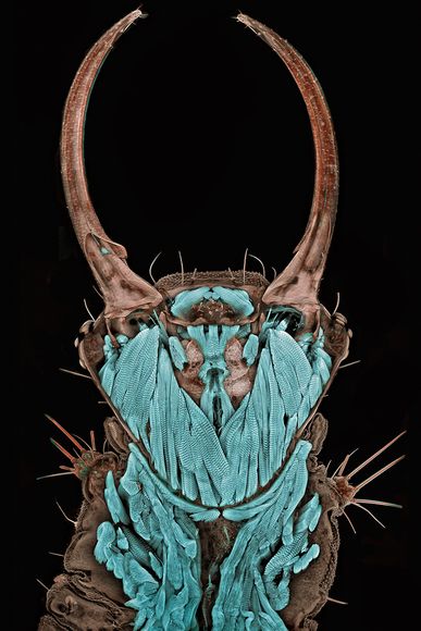
Lacewing Portrait: photo of Igor Siwanowicz, Max Planck Institute of Neurobiology / Nikon Small World.
Exaggerated 20 times, the lower jaw of the larvae looked quite fierce. The photo won at the Small World Microphotography Competition in 2011.
The description of very small parts of insects began when Igor Siwanowicz's hand was bitten by them, prompting the photographer and other biologists to take the insect he kept in his pocket into the test tube.
Returning to the lab, in Madison, Wisconsin, Siwanowicz carefully pinned and stained Lacewing who had died for his photography job, which was no simple task, as the length of the head was only 1.3 millimeters.
'My art causes discord for viewers, the contradiction between an insect's perception of ugliness and its inner beauty ,' he said.
Sponsored by Nikon, the Small World competition honors images taken with light microscopes 'successfully in introducing the delicate combination of scientific and artistic techniques '.

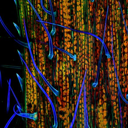
Grass leaf: photo by Donna Stolz, Pittsburgh University / Nikon Small World.
The microscopic strands of a large grass leaf were expanded 200 times in a fluorescent microphoto, making Donna Stolz from the University of Pittsburgh win the second place of the competition.
This photo is one of 92 winning photos at this year's microscope photo contest, candidates from 70 countries.

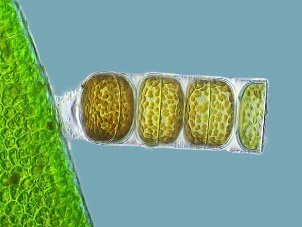
Algae: photo by Frank Fox, Trier / Nikon Small World University.
A living specimen of the Melosira moniliformis (right) is magnified 320 times in microphoto by Frank Fox, University of Trier, Germany.
Top photos from the competition Nikon Small World 2011 photos will be displayed in a colorful calendar and in a travel museum in the United States.

Fourth position: Lichen: photo by Robin Young, British Columbia University / Nikon Small World.
They may look like lizard feet, but these colorful models come from a branch of liverwort - a plant species. Robin Young exaggerated liverwort 20 times to win the award for this photo.

Microchip surface: picture of Alfred Pasieka.Nikon Small World
Looking like a maze, this 3D image is Microchip's surface magnified 500 times by a German, named Alfred Pasieka. As in the past year, a panel of journalists and scientists has won at Small World 2011.

Membrane cracks on solar cells: photo by Dennis Callahan, California Institute of Technology / Nikon Small World.
This crack was magnified 50 times, solar cells became abstract art in the sixth ranked photo, performed by Dennis Callahan from California Institute of Technology. The membrane containing gallium-arsenic, a mixture of gallium and arsenic elements forms semiconductors in solar energy cells.

The nerve structure of the mouse: a picture of Gabriel Luna, UCSanta Barbara / Nikon Small World.
The image is magnified 40 times with a laser scanning microscope, showing the complex network of nerves in the retina.

Colorful granulit: photo by Bernardo Cesare, Padova University / Nikon Small World.
A picture with bold, vivid colors of graphitxengranulit, a deformed rock consisting mainly of feldspar and quartz. Bernardo Cesare of Padova, exaggerated the stone - collected from Kerala, India 2.5 times and won the 8th place of the competition.

Marine Copepod: photo by Jan Michels, Kiel University / Nikon Small World.
This is a small marine crustacean, which is one of the most common species in the multicellular world under the ocean. Jan Michels, Kiel University in Germany took the abdomen and exaggerated 10 times.

Water fleas: photos of Joan Röhl, Biochemistry and Biology Institute / Nikon Small World
Exaggerated 100 times, the inner workings of a fresh water flea (approximately 5 millimeters in length, about 0.2 inches) appear in the painting by German Joan Röhl.
- Best micro photos in 2010
- Photos of the most beautiful micro-world in 2010
- Micro photos of meteorites reveal a lot of information
- Introduce robot can learn as human brain
- Detecting micro-plastic particles in rainwater, not only lies in the sea
- Exaggerated photos won in 2008 (Part 1)
- Exploding the micro grid market
- Each person swallowed at least 50,000 micro-plastic beads every year
- The photos imagined photoshop but 100% real
- Plants under a microscope
- Put the micro algae robots into the body to spy on and treat cancer
- Corals reject shrimp eggs, choose to eat micro-plastic seeds
 The 11 most unique public toilets in the world
 Explore the ghost town in Namibia
 Rare historical moments are 'colored', giving us a clearer view of the past
 The world famous ghost ship