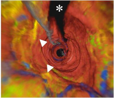
The first 3-D image in human arteries
A team of researchers said that the wall in the coronary arteries in the human body for the first time is shown in 3-D images.
These images allow cardiac surgeons to see the inside of the patient's arteries more clearly and check for areas of infection inside the fibrous patches, the cause of a heart attack. .
Researcher Dr. Gary Tearney, also an assistant professor of pathology at Harvard Medical University, said: "It proves that people have the technical ability to help cardiologists. can look deep inside coronary arteries. The abundance of information helps us to achieve a clear improvement in our ability to understand coronary diseases and allow cardiologists to remove sequelae before it spread to a more serious city. "

3-D images of coronary arteries.(Photo: Massachusetts General Hospital)
The research team at the Massachusetts Institute of Wellman Medicine Center (MGH) developed a visual frequency device to identify human arteries in 3-D form. This device, called OFDI for short, can see more than 1,000 points of arterial tissue at the same time (previous devices can only calculate one point in a cell). Naturally they will probe throughout the coronary arteries, the wavelength will spread and reflect, quickly allowing the acquisition of the required data to create super-sophisticated image details.
Not only does this device provide 3-D images in a second, the device's extremely fast speed also reduces the prescription signs in the blood, causing infections of previous technical devices.
Recent research, led by Professor Tearney, collected information, detailed images of arteries from patients - lipid fluids or calcium levels and immune cells that could indicate a place inflammation. And that allows you to delve into and see inside the human body with more detail. The more horizontal and vertical images of each vascular array are actually related to plaque causing degeneration and the cause of heart attacks.
Tearney and his colleagues noted that this work was successful thanks to the support of the majority of patient groups, and the need to proceed most quickly to bring equipment into operation soon. in real life to reduce the time and difficulties for cardiologists. Combining OFDI devices with cardiovascular ultrasound can help greatly support the limitations of technology, which we have been helpless for penetrating into tissue cells for a long time.
The next step is to make sure that this technique is widely used, potentially becoming a commercial device.
- English students solve the problem
- New injection for cancer treatment
- Wireless devices move in the arteries
- Interesting discovery of human circulatory system
- Food for people infected with fibrosis
- For the first time, the image of ovulation is recorded
- Computers have been able to read human thoughts almost in real time
- Heart failure, coronary arteries increase
- New technique of coronary artery: Five hours of surgery, ten years of health
- Create a '4D' image of the human body
- Both blood pressure measurements help detect heart disease early
- A close-up picture of the original human image
 Daily use inventions come from universities
 Special weight loss device helps prevent appetite
 8 inventors were killed by their own inventions
 Iran invented a motor car powered by water
 Wireless devices move in the arteries