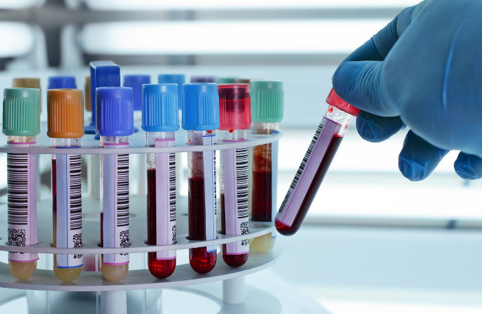
Instructions on how to read blood test results
How to read the results of blood or urine tests is what you need when you have a physical exam because doctors still or often appoint patients to do blood tests and urine tests. However, if the doctor does not explain it, you cannot understand what the indicators on the test result mean. Please follow the article below to learn how to read the blood test results.
I. COMPONENTS OF BLOOD FORMULA
- WBC (White Blood Cell - Number of white blood cells in a blood volume):
The value is usually in the range of 4,300 to 10,800 cells / mm 3 , which is equivalent to the number of leukocytes in international units of 4.3 to 10.8 x 10⁹ cells / l.
Increased in inflammation, malignant blood disease, leukemia .; reduction in aplastic anemia, vitamin B12 deficiency or folate, bacterial infection .

Values usually range from 4,300 to 10,800 cells / mm3.
- RBC (Red Blood Cell - Number of red blood cells (or erythrocyte count) in a blood volume):
The value is usually between 4.2 and 5.9 million cells / cm 3 , equivalent to the number of erythrocytes in international units of 4.2 to 5.9 x 10¹² cells / l.
Increased in dehydration, erythrocytosis; decrease in anemia.
- HB or HBG (Hemoglobin - Amount of hemoglobin in a blood volume):
Hemoglobin is a type of protein molecule in red blood cells that carries oxygen and produces red blood cells.
Values vary by gender, usually between 13 and 18 g / dl for men and 12 to 16 g / dl for women (8.1 to 11.2 millimole / l, respectively, and 7.4 - 9.9 millimole / l).
Increased in dehydration, heart disease and lung disease; decrease in anemia, bleeding and reactions that cause hemolysis.
- HCT (Hematocrit - Ratio of erythrocyte volume on total blood volume):
Value varies by gender, usually between 45 and 52% for men and 37 to 48% for women.
Increased in allergic disorders, erythrocytosis, smoking, chronic obstructive pulmonary disease, coronary artery disease, in the mountains, dehydration, hypotension; reduction in blood loss, anemia, pregnancy.
- MCV (Mean corpuscular volume - Average volume of a red blood cell):
This value is taken from HCT and the number of red blood cells. Normal values range from 80 to 100 femtoliter (1 femtoliter = 1/1 million liters).
Increased in vitamin B12 deficiency, folic acid deficiency, liver disease, alcoholism, hyperemia, hypothyroidism, bone marrow aplasia, bone marrow fibrosis; reduction in iron deficiency, thalassemia syndrome and other hemoglobin diseases, anemia in chronic diseases, anemia of erythrocytes, chronic renal failure, and lead poisoning.
- MCH (Mean Corpuscular Hemoglobin - Average number of hemoglobin in a red blood cell):
This value is calculated by measuring hemoglobin and the number of red blood cells. Normal values range from 27 to 32 picograms.
Increase in normal erythrocyte anemia, severe hereditary circular erythrocyte, the presence of cold agglutination; reduction in the initiation of iron deficiency anemia, general anemia, regenerative anemia.
- MCHC (Mean Corpuscular Hemoglobin Concentration - Average concentration of hemoglobin in a blood volume):
This value is calculated by measuring the value of hemoglobin and hematocrit. Normal values range from 32 to 36%.
In sharp anemia: normal red blood cells, severe hereditary circular erythrocytes, the presence of cold agglutination factors.
In renewable anemia: may be normal or decrease due to reduced folate or vitamin B12, cirrhosis, alcoholism.
- PLT (Platelet Count - Platelet Count in a blood volume):
- Platelets are not a complete cell, but fragments of cytoplasm (a cell component that does not contain the nucleus or stem of the cell) from cells found in the bone marrow.
- Platelets play a vital role in blood clotting, with an average life expectancy of 5 to 9 days.
- Values usually range from 150,000 to 400,000 / cm 3 (equivalent to 150 - 400 x 109 / l).
- Too low platelet counts will cause blood loss. A high platelet count will form a blood clot, impede blood vessels, leading to stroke, myocardial infarction, pulmonary embolism, blood vessel blockage .
- Increased in bone marrow proliferation disorders, idiopathic thrombocytopenia, bone marrow fibrosis, post-bleeding, postoperative splenectomy ., leading to inflammatory diseases.
- Reduction in bone marrow suppression or replacement, chemotherapy, splenic hypertrophy, scattered intravascular coagulation, platelet antibodies, purpura after blood transfusion, and thrombocytopenic thrombocytopenia Infant.

Lymphocyte helps the body fight infections.
- LYM (Lymphocyte - Lymphocytes):
Lymphocyte helps the body fight infections. There are many causes to reduce lymphocytes such as immunity, HIV / AIDS, TB, malaria, leukemia, lymphoma .
Normal values range from 20 to 25%.
- MXD (Mixed Cell Count - blood cell mixing ratio):
Each type of cell has a certain percentage of blood. MXD varies depending on the increase or decrease in the ratio of each cell type.
- NEUT (Neutrophil - Neutrophil ratio):
Normal values range from 60 to 66%. High incidence indicates blood infection.
Increased in acute infection, acute myocardial infarction, stress, cancer, myeloid leukemia; reduction in viral infection, aplastic anemia, immunosuppressive drugs, radiation therapy .
- RDW (Red Cell Distribution Width):
This higher value means that the distribution of red blood cells changes more and more. Normal values range from 11 to 15%.
Normal RDW and:
- MCV increases, encountered in: aplastic anemia, before leukemia.
- MCV is normal, encountered in: anemia in chronic diseases, blood loss or acute hemolysis, enzyme disease or anemia without anemia.
- MCV reduced: anemia in chronic diseases, heterozygous thalassemia
RDW increases and:
- MCV increases: vitamin B12 deficiency, folate deficiency, immune hemolytic anemia, cold agglutination, chronic lymphocytic leukemia.
- Normal MCV: early iron deficiency, early stage vitamin B12 deficiency, early folate deficiency, anemia caused by globin disease.
- MCV decreases: iron deficiency, red cell fragmentation, HbH, thalassemia.
- PDW (Platelet Disrabution Width -: Platelet Disrabution Width):
Normal values range from 6 to 18%.
Increase in lung cancer, sickle cell disease, gram positive, gram-negative sepsis; Reduce in alcoholism.
- MPV (Mean Platelet Volume - The average volume of platelets in a blood volume):
Normal values range from 6.5 to 11fL.
Increased in cardiovascular disease, diabetes, smoking, stress, poisoning by thyroid .; reduction in anemia due to aplasia, giant erythrocyte anemia, cancer chemotherapy, leukocytosis .
- P-LCR (Platelet Larger Cell Ratio - Large platelet ratio):
Normal values range from 150 to 500 G / l (G / l = 109 / l).
II. HOW TO READ THE BLOOD RESULTS

GLUCOSE is blood sugar.The normal limit is from 4.1 to 6.1 mnol / l.
1. GLU (GLUCOSE): Blood sugar. The normal limit is from 4.1 to 6.1 mnol / l. If the limit is exceeded, increase or decrease blood sugar. Above the limit is a person at high risk for diabetes.
2. SGOT & SGPT: Liver enzyme group
The normal limit is from 9.0-48.0 with SGOT and 5.0-49.0 with SGPT. If this limit is exceeded, the hepatotoxicity function of the hepatocellular cells decreases. Limit food intake, drinking water makes the liver difficult to absorb and affect liver function such as:
Animal fats and alcohols and carbonated drinks.
3. BLOOD CLASS: Includes CHOLESTEROL, TRYGLYCERID, HDL-CHOLES, LDL-CHLES
The normal limit of these group elements is as follows:
- Normal limits range from 3.4-5.4 mmol / l with CHOLESTEROL.
- Normal limits range from 0.4-2,3 mmol / l with TRYGLYCERID.
- Normal limits range from 0.9-2.1 mmol / l with HDL-Choles.
- Normal limits range from 0.0-2.9 mmol / l with LDL-Choles.
If one of the above factors exceeds the permitted limit, there is a high risk of cardiovascular disease and blood pressure. Particularly HDL-Choles are good fats, if high it limits blood clots. If the CHOLESTEROL is too high with high blood pressure and high LDL-Choles, the risk of stroke and stroke is very high. Avoid eating foods high in fat and cholesterol such as animal viscera, poultry eggs, shrimp, crabs, beef, chicken skin . Enhancing sports. Drink more garlic alcohol and monitor blood pressure regularly.
4. GGT: Gama globutamin, is an immune factor for liver cells. Normally, if liver function is good, GGT will be very low in the blood (From 0-53 U / L). When liver cells have to work excessively, the ability of liver to be toxic is reduced, the GGT will increase -> Reduce the resistance and immunity of liver cells. Easy to lead to liver failure. If people with SVB in the blood and GGT, SGOT & SGPT have increased, it is necessary to use liver cells and absolutely no alcohol or the risk of VGSVB is very high.
5. URE (blood urea): is the most important degradation product of protein that is excreted by the kidneys.
Normal limit: 2.5 - 7.5 mmol / l.
6. BUN (Blood Urea Nitrogen) = urea (mg) x 28/60; Unit conversion: mmol / lx 6 = mg / dl.
- Increase in: kidney disease, high protein intake, fever, infection, urinary obstruction .
- Reduce in: low protein intake, severe liver disease, depletion .
BUN: is the nitrogen of urea in the blood.
Normal limit 4.6 - 23.3 mg / dl. -> Bun = mmol / lx 6 x 28/60 = mmol / lx 2.8 (mg / dl).
- Increase in: kidney failure, heart failure, high protein intake, fever, infection .
- Reduce in: eating less protein, severe liver disease .
7. CRE (Creatinine): is the product of excretion of muscle degradation in muscle creatin, the amount of formation depends on muscle mass, is filtered through glomerular & excreted in the urine; also the most stable protein component regardless of diet -> has a value for determining glomerular function.
Normal limit: male 62 - 120, female 53 - 100 (unit: umol / l).
- Increased in: kidney disease, heart failure, diabetes, idiopathic hypertension, acute MI .
- Reduce in: pregnancy, eclampsia .
8. URIC (Uric Acid = urat): is a metabolite of purine base (Adenin, Guanin) of DNA & RNA, mainly discharged through urine.
Normal limit: male 180 - 420, female 150 - 360 (unit: umol / l).
Increase in:
- Originally: due to increased production, due to reduction (spontaneous) -> related enzymes: disease of Lesh Nyhan, Von Gierke .
- Secondary: due to increased production (myeloma, psoriasis .), due to reduced excretion (kidney failure, medication, atherosclerosis .).
- Gout (gout): hyperuricemia / blood may be accompanied by tophi nodules in the joint & kidney urate stones.
Reduce in: Wilson disease, hepatocellular injury .
9. FREE RESULTS
- Anti-HBs: Antibody against hepatitis B virus in the blood (ATTENTION <= 12 mUI / ml).
- HbsAg: Hepatitis B virus in the blood (CALCULATION).
- Instructions on how to read urine test results
- The things doctors won't reveal in your blood test
- How to use a pregnancy test stick and how to read the most accurate pregnancy test results
- Summary of the best news on September 2
- Things to know about general blood tests
- Best news aggregation for the week of October 2
- Normal indicators when evaluating a semen test
- Summarize the best news from August 3
- Quick and accurate blood tests with SpinChip
- How to read the results of eye test results?
- What blood tests can you know about?
- Automatic robotic blood collection system
 13 causes of non-itchy rash
 How the mouse with human ears changed the world?
 The truth about 'fried rice syndrome!
 What is dental implant?