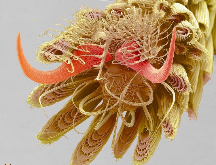
The photo revealed the mosquito's feet and scales
Snapshot enlarges 800 times the foot of the mosquito attracts a lot of attention on social networks with a structure covered with feathers and scales.
A photograph of a mosquito footage from a scanning electron microscope of Steve Gschmeissner photographer fevered on social network Reddit with more than 32,000 likes, Live Science reported. First appeared in the Royal Photographers Association's 2016 International Science Photo Contest, the photo was shared many times on social networks from then on, possibly due to its complexity, according to Gschmeissner.

Mosquito foot photograph of Steve Gschmeissner.(Photo: Live Science).
In the picture is the end of a mosquito's leg, including the cork, a lot of small scales and a cushion of hairy feet. According to the description of Gschmeissner's image, these scales cover the mosquito's body but are especially thick near the foot, which protects and allows mosquitoes to land on the water, where they lay eggs."Insects are great because they have all kinds of tiny details , " Gschmeissner said.
Gschmeissner has a degree in zoology and dozens of years of experience in cancer research and scanning (SEM) at the Royal University of Surgery in London, England. SEM glasses emit a beam of electrons through the object to capture. The object is coated with gold so that electrons disperse on the surface, giving information about the shape. Due to the use of electrons, SEM has a very high resolution and can even capture nanoscale structures. In the picture, the mosquito's foot was enlarged 800 times.
Gschmeissner has explored the diversity of the microscopic world with this technique. He photographed invisible human neighbors, such as the bacteria found on dish towels and cell phones, the bacteria that live in the human mouth and kidneys, or the termites that reside on eyelashes, pollen and sign germ.
- Strange feet in the world
- 18 interesting things about dog feet
- Detection of wound healing of collagen in fish scales
- 7 facts make you fall back on the ivory pearl legs of cat bosses
- The scales are as hard as the armor of an Amazon arowana
- How to identify dirty chicken feet to avoid poisoning
- Treat cold hands and feet
- This is why eating chicken feet is good for health
- The huge foot of the large legged suspect
- Bulletproof material new varieties of fish scales?
- Guilty feet
- Robot feet simulate perfect human movement
 Why do potatoes have eyes?
 'Tragedy' the world's largest carnivorous life: Death becomes ... public toilet
 Tomatoes were once considered 'poisonous' for 200 years
 Detecting microscopic parasites on human face