50 years ago the first CT scanner let doctors see inside the skull
The possibility that precious items are hidden in secret rooms can spark a lot of imagination.
In the mid-1960s, British engineer Godfrey Hounsfield wondered if it was possible to detect such mysterious regions inside the Egyptian pyramids by capturing cosmic rays passing through unobserved voids. Okay.
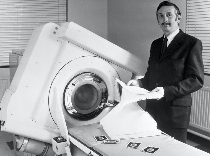
Godfrey Hounsfield stands by an EMI-Scanner in 1972. (Image: Getty Images)
For years, Hounsfield struggled with the idea of 'looking inside a sealed box without opening it'. He eventually determined how to use high-energy rays to reveal what is invisible to the naked eye.
Hounsfield succeeded with inventing a way to look inside the skull and take pictures of the human brain. He had never been to Egypt to explore the secret chambers of the pyramids, but that invention brought him to Stockholm to receive the Nobel Prize and to Buckingham Palace to receive a Knighthood.
Creative Electrical Engineer
Hounsfield was born in Sutton-on-Trent, Nottinghamshire, England in 1919. From an early age, he enjoyed tinkering with electrical and mechanical devices. At the start of World War II, Hounsfield joined the Royal Air Force, but not as a soldier. Hounsfield is a 'witch' with electrical machines, especially with the newly invented radar to help pilots better find their way home in dark and cloudy skies.
After the war, Hounsfield continued his studies and earned a degree in engineering. He worked for EMI, which was then best known for its Beatles recording and album sales, but started off focusing on electronics and electrical engineering.
Hounsfield's natural talent made him the leader of the most advanced mainframe computer group in Britain. But by the 1960s, EMI wanted to get out of the computer market, and they didn't know what to do with a brilliant, eccentric engineer.
During a vacation, Hounsfield reflected on his future and what he could do for the company. He saw a doctor who complained about the poor quality of brain X-rays. The X-ray showed only the marvelous details of the bones, but the brain remained an amorphous mass of tissue on film. It all looks like fog. This made Hounsfield think of his old idea of "finding hidden structures without opening the box".
Unveil the unseen
Hounsfield has come up with a new way to deal with the problem of imaging the inside of the skull.
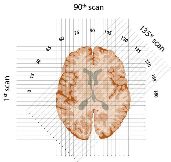
X-rays pass through each "slice" of the brain, oriented at each degree in an arc from 1 to 180 degrees in a semicircle.
First, conceptually, he divides the brain into consecutive slices - like a loaf of bread. He then plans to shine a series of X-rays through each layer, repeating this for each degree of the semicircle. The intensity of each beam will be recorded on the opposite side of the brain - and stronger beams indicate they have passed through less dense material.

Calculating the intensity of each X-ray as it passes through the object, and using an impressive algorithm, it is possible to create an image of the inside of the "closed box".
Finally, in perhaps his most ingenious invention, Hounsfield created an algorithm to reconstruct an image of the brain based on all these layers. Using one of the fastest new computers of the time, he was able to calculate the value for each 'small box' of each brain layer.
But there was a problem: EMI didn't enter the medical market and didn't want to jump in. The record company still allowed Hounsfield to research its products, but with a meager amount of funding. He was forced to rummage through scrap bins of research facilities and assemble a rudimentary scanner, small enough to fit on a dining table.
Even if the scanner succeeded in scanning inanimate objects, and later Kosher cow brains, EMI's funding would have gone nowhere. Hounsfield needs outside funding if it wants to build a brain scanner.

Schematic of the CT scanner is included in Hounsfield's US patent.
Hounsfield was a brilliant inventor, but not a good diplomat. Luckily he had a sympathetic boss, Bill Ingram, who saw value in Hounsfield's proposal and fought with EMI to allow the project to continue.
Success with brain tumor detection
Bill Ingram knows that funding would ideally need to come from the government, on the grounds that the UK Department of Health and Social Security could be a customer to buy equipment for hospitals. And miraculously, Ingram sold them 4 scanners even before they were built. At this time, Hounsfield organized a team of engineers, focused on building a brain scanner that was safe and effective for humans.
The team installed a full-size scanner at London's Atkinson Morley Hospital, and on October 1, 1971, they scanned the brains of their first patient: a middle-aged woman with signs of a tumor. Brain.
It's not a quick procedure – it takes 30 minutes for the scanner, a drive-through with tapes across the city; 2.5 hours of data processing on the EMI mainframe and imaging with the Polaroid before returning to the hospital.
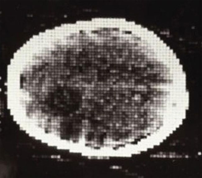
In the first clinical CT scan, the brain tumor is visible as a darker spot.
And doctors saw in the patient's left frontal lobe a cyst the size of a plum. With that invention, all other methods of brain imaging became obsolete.
Millions of CT scans per year
EMI, which had no experience in the medical market, suddenly held a monopoly on a medical device that was in great demand. The scanner invented by Hounsfield went into production and sold very well. But within the next five years, larger, more experienced companies with more research capabilities like GE and Siemens produced better CT scanners and dramatically increased sales. The EMI must then leave the market.

Swedish King Carl Gustaf presents the Nobel Prize to Hounsfield in Stockholm on December 11, 1979. (Image: Getty Images)
Hounsfield's invention transformed the world of medicine . He was awarded the Nobel Prize in Medicine in 1979 and knighted by the Queen of England in 1981. Hounsfield continued with his inventions until his death in 2004, at the age of 84.
In 1973, Robert Ledley, an American, developed a full-body scanner that could image other organs, blood vessels and, of course, bones. Modern scanners are faster, provide better resolution, and most importantly, patients undergo the procedure with less radiation exposure.

Modern CT scans provide much higher resolution images than Hounsfield's original 1971 "slice" of the brain.
By 2020, technicians are performing more than 80 million CT scans annually in the US. Some doctors consider this number to be excessive and perhaps a third is unnecessary. While that may be true, CT scans have benefited the health of many patients around the world, helping to identify tumors and determine if surgery is needed. They are especially useful in quickly finding damage inside the brain after an accident.
And recall Hounsfield's idea of pyramids. In 1970, scientists placed a cosmic ray detector in the lowest chamber in the Pyramid of Khafre. They concluded that no mysterious room existed inside this pyramid. In 2017, another research team placed a cosmic ray detector at the Great Pyramid of Giza and found a hidden but inaccessible room. And it is unlikely that this place will be discovered any time soon.
- 3D scanner made in Vietnam
- 3D scanner launched
- Russia invented pocket-sized anti-terrorism scanner
- The surgery for piercing the skull was 3,000 years ago
- Mysterious skull surgery 2300 years ago
- Netherlands: Successfully implanted 3D skull for a woman
- Detecting skulls dating to 430,000 years old
- New discoveries in Peru reveal the possibility of the Inca skull surgery
- The world's first full-body 3D scanner
- Unique pen capable of scanning documents and recording
- Found 16,000-year-old human skull nearly intact
- Monkey skull fossils 20 million years ago have the potential to reveal the evolution of the human brain
 Biography of hero Vu A Dinh
 History of hematology
 Who is Mr. Tam Da 'Phuc-Loc-Tho' and what does it mean?
 Unbelievable facts about the history of the oil and gas industry: Gasoline used to be cheaper than water, so abundant that it had to be dumped into the river...