Retinoblastoma: Causes, symptoms, diagnosis and treatment
Retinal cancer is a life-threatening disease, but if treated properly and promptly, it rarely causes death.
What is retinoblastoma?
Retinoblastoma is also known as retinal cancer is a malignant eye disease seen in young children. This is a dangerous disease that not only destroys the visual function of the eye but can also be life-threatening. The tumor may develop in one or two eyes. The disease may be caused by genetic or sporadic factors. Genetic retinoblastoma of children can also cause other types of cancer.
Most patients are diagnosed before age 2 but can be detected at birth and in adults older than 52 years.
If the disease is detected and treated early, it can be cured with a 5-year survival rate of up to 90-95% and keep the eyeball high.
Retinal cancer is a malignant eye disease seen in young children.
The cause of retinoblastoma is due to the genetic abnormality or not.
- Retinoblastoma is related to family factors: 6%. The disease usually manifests early when the baby is a few months old, often with both eyes. Your child may have another type of cancer.
- Retinoblastoma is not related to family factors: accounts for 94%. The cause of gene mutation in which 80% has no genetic ability and 20% may be hereditary.
Symptoms of retinoblastoma
Symptoms of retinoblastoma depend on the size of the tumor, the stage of the disease and the complications of the tumor in the eye. Some cases are detected by an eye examination for premature babies or school children.
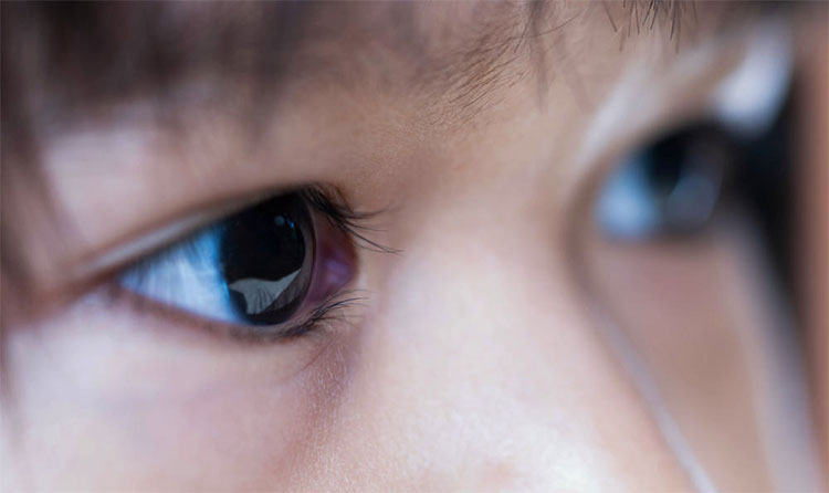
- Signs of white pupil: More than 50% of the disease is detected from white pupil signs. Signs can be described by different words such as 'cat eyes', 'beast eyes' . When looking at the eyes of children, there will be light, one or two pupils may be white or yellow when taking photos of the session. I used the flash at night. This sign is particularly clear at night or in a dark room because then the pupil will relax.
- Signs of strabismus: More than 30% of childhood retinoblastoma is detected from this sign.
- Visual acuity decreased: 8% of cases were detected by this sign.
- Other signs: redness and soreness of the eyes due to glaucoma, uveitis, ocular cell inflammation, pre-haemorrhage .
- Late stage tumors can progress to the back of the eyeball such as invading the optic nerve, spreading the eye socket and distant metastasis. Invasive tumor through the optic nerve or subarachnoid subarachnoid. The tumor can spread to the skull, spinal cord and lymph nodes. Since then the tumor follows blood vessels, lymphatic metastases far away from the organs in the body.
Clinical forms of childhood retinoblastoma include:
- Retinoblastoma on one side of the eye: 75% of cases occur, children aged 2-4 years.
- Retinoblastoma of the bilateral eyes: 25% of children younger than 14-16 months of age are inherited in 40%.
- Trigeminal retinoblastoma: retinoblastoma and retinoblastoma, accounting for 3-9%. The disease has a very poor prognosis. Most children die within 35 months.
Stages of retinoblastoma:
Retinoblastoma is staged according to S. Jude Children's Research into 4 stages:
- Stage 1: U (one drive or multiple drives) is still localized in the retina.
- Stage 2: U (one drive or multiple drives) spread outside the retina but is limited in the eyeball.
- Stage 3: U invasion of the eyeball, intracranial metastasis.
- Stage 4: Having metastasis far away from blood sugar (internal organs, bones, bone marrow .).
Subjects at risk of retinoblastoma:
- Young children, especially under 2 years old
- Children have abnormal eye symptoms such as strabismus, red eye, eye hemorrhage, cat eye image .
- Children with sick family members
Prevention of retinoblastoma:
- There are no specific preventive measures
- The best way is to have an eye exam for children when there are any signs of suspicion
- Examination and screening for children with family factors
Tests in the diagnosis of retinoblastoma include:
- Brain scans: 75% of cases of calcification in the eye and can determine the tumor invasion of the tumor
- Ultrasound: especially useful in case of cloudy cloudy like cloud cover
- Computerized tomography: advanced techniques to determine the calcification and pre-assessment of treatment of disease in the optic nerve, in the eye and in the brain
- Determination of LDH in vitreous: Elevated LDH is found in more than 90% of retinal cancer patients
- Elevated levels of CEA and AFP in the blood then decline to normal levels after removal of the eyeball.
- Cerebrospinal fluid tests evaluate tumor invasion into the CNS
- Disease surgery: not a mandatory test when diagnosing retinoblastoma because of diagnosis mainly by non-invasive eyeball methods. Pathology has postoperative value to evaluate the extent of the tumor invasion.
Treatment measures for retinoblastoma
The combination of surgical, chemical and radiotherapy treatments achieved satisfactory results with the overall survival rate after 5 years with an intraocular tumor of 90%, external invasive tumors of 10% . The current treatment trend is to live more with the patient's vision.
Surgery to remove the eyeball
Point:
- Large tumor (> 60% of eyeball volume)
- The patient has no vision
- The tumor invaded the nervous market, invading the room
- Failure of previous conservative treatments
After surgery can be chemical or radiotherapy auxiliary in cases of invasive iris, ciliary folds, choroid .
After an eyeball, a fake eye can be fitted to the child.
Surgical complications: spreading cancer cells into the eye.
Radiation
Indicated in the case of large tumors on either side, sowing seeds into the lens, tumors are near the optic nerve
Complications of radiation: retinal damage, optic nerve, lacrimal glands and vitreous lens
The second cancer that appears after radiation is also a complication, especially in patients with genetic retinoblastoma.
Freezing laser
Applicable to tumors of small size, width
Direct treatment within the tumor, freezing the blood vessels that supply the tumor
Chemistry
Chemicals are used in cases such as:
- Polyps invade> 25% of the region's retina without radiation therapy
- All large tumors are not removed
- U invades the cornea
- Lesions spread outside the eyeball.
Bad prognostic factors of the disease include:
Delayed diagnosis of diseases greater than 6 months.
A history of intraocular surgery that is capable of seeding into glass or spreading malignant cells out of the eye.
The patient has cataracts.
Radiotherapy using external rays can lead to secondary cancers, especially in patients with hereditary retinoblastoma.
Tumors that invade the choroid, optic nerve, or eye socket increase the risk of metastasis.
- The risk of spreading into the optic nerve: exogenous development (eg, from the outer layers of the retina towards the choroid), increasing intraocular pressure and thickness of ≥ 15 mm.
- Risk of spread into the choroid: increased intraocular pressure; Renewal blood vessels iris.
The prognosis depends on the condition, location and degree of recurrence of the tumor.
- New findings about Alzheimer's disease help early diagnosis and increase healing
- Dangerous cancer - cause and prevention
- A breakthrough in cancer diagnosis and treatment
- Irritability: Causes, symptoms, diagnosis and treatment
- Detection of cancer, asthma, diabetes and many other diseases through ... fingerprints
- The new algorithm can predict disease ... 48 hours before symptoms appear
- Where should the Zika virus be tested in Vietnam?
- New method of diagnosis and treatment of breast cancer
- Detect eye cancer in young children by turning on the flash when taking photos
- Allergic rhinitis: Causes, symptoms and treatment
- Manufacturing equipment for early diagnosis of throat cancer
- Ultrasound can early diagnose uterine cancer
 Why is Australia the country with the highest cancer rate in the world while Vietnam ranks 100th?
 New drug causes cancer to 'starve'
 Common cancers in men
 America's incredible discovery: The most feared cancer cell is love