The most breakthrough scientific images in 2009
The Wellcome Images award is given by the Wellcome Trust, a humanitarian organization dedicated to funding health studies. Over the past 10 years, this organization has awarded awards for groundbreaking photographs in the field of medical research, social history, health care and biology.
Here are some photos of the 2009 Wellcome Images award.
1. The image is taken through a microscope of seeds of natural flowers - a flower that is likened to the bird's paradise. This flower is native to Africa. Their flowers often have two characteristic colors: orange and blue. The first image of the celestial flower seeds was pictured in the watercolor painting by painter Annie Cavanagh, but was later captured by photographer Dave McCarthy as shown below. (Photo: Annie Cavanagh and Dave McCarthy)

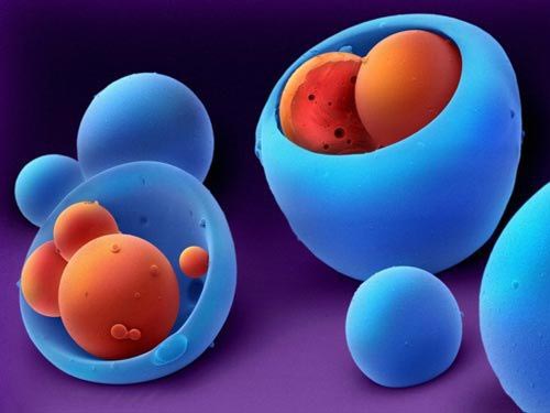
2. Copolymers can be used in microparticles or in the production of microorganisms. Pilime polymers that do not decompose or decompose slowly in acidic environments can be used as covers for capsules. This helps reduce the number of times a patient has to take medication for a day.
In the picture, orange beads are drugs that contain prednisolone, which she uses for arthritis. They are surrounded by a co-polymerization layer.(Photo: Annie Cavanagh)

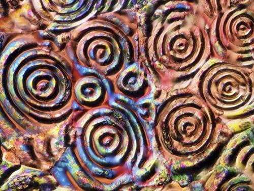
3. This is a picture of a crystal aspirin taken through a microscope. Aspirin is not only used as an analgesic and anti-inflammatory, but also used for anti-coagulation. (Photo: MI Walker)

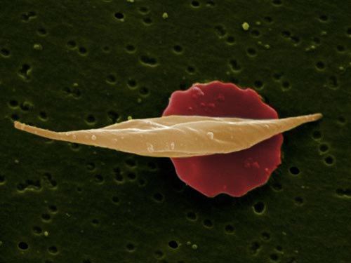
4. This is an image of two red blood cells. This image shows two red blood cells. A normal red blood cell has a red circle, but when the red cells are deformed into sickle cell red blood cells, they reduce the ability to transport the substance. High sickle cell count will reduce our body's resistance to malaria. (Photo: Jackie Lewin) .

5. The photo of a feather is shaped like a finger on the small intestine wall of the mouse. (Photo: Paul Appleton) .

6. Professor Harold Kroto, who won the 1996 Nobel Prize in Chemistry for his work on carbon structure and fullerine technology. This research was done by Robert Curl and Richard Smalley. In the picture is Professor Harold Kroto at the lab the day after the Nobel Prize for his work was published. (Photo: Anne-Katrin Purkiss)

7. This is an image of capillaries (small blood vessels). They act as a bridge between veins and arteries. (Photo: Spike Waker)

8. This is a digitally reconstructed photo from a pencil drawing of the structure of the heart on the human body. (Photo: Bill Mccokey) .

9. The image of the rat liver was taken through an electron microscope. Sinus blood vessels have a very important role to help the liver produce bile. The image also contains blue lines, which are bile ducts to the intestines to help digest food. (Photo: Jackie Lewin) .

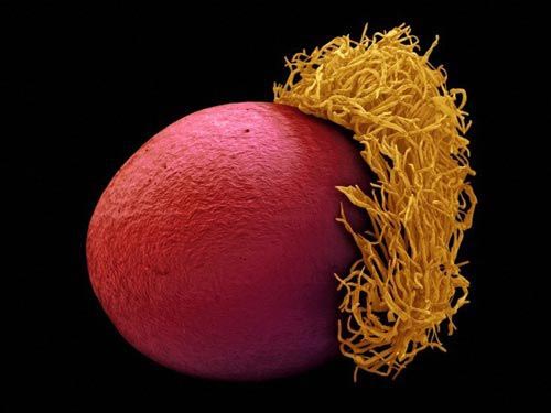
10. This is an image of in vitro fertilization. The sperm are trying to break the protective shell in the eggshell. If sperm passes through this ' barrier ', a living body will be formed. (Photo: Spike Walker) .

11. Images of lung cancer cells taken from electron microscopy. Purple cancer cells grow in clusters on the cell surface of the lungs. (Anne Weston) .

12. A group of scientists have built DNA structures using 3D graphics technology. (Photo: Oliver Burston)

- The biggest scientific breakthrough in 2010
- The 2018 Breakthrough Prize scientific awards are now available
- Collect 3-dimensional images of flu viruses
- Breakthrough research proves that two men can have children together
- Scientists launch a million-dollar project to
- The work of a breakthrough Vietnamese graduate student in Australia
- 10 most viewed scientific photos in 2012
- 10 scientific breakthroughs in 2014
- Breakthrough scientific findings in 2016
- The first image of the black hole was awarded a $ 3 million 'Science Oscar'
- The discovery of the neutron star collision is 'breakthrough' of 2017
- The Nobel Prize for Breakthrough by the
 The 11 most unique public toilets in the world
 Explore the ghost town in Namibia
 Rare historical moments are 'colored', giving us a clearer view of the past
 The world famous ghost ship
 Photos capture Shanghai's golden age, every frame is as beautiful as a movie
  Revealing a series of rare photos recording little-known things in history
  Top 9 Gods in 'Greek Mythology'
  Photographs of Chinese Women 150 Years Ago
  Leaked photos of Leonardo da Vinci and Mona Lisa, experts join in to find surprising clues
  Birds that don't look at each other win prestigious European nature photo award