Giant viruses infect cells like?
The researchers found giant viruses invading cells through a special star-shaped structure on the crust.
In a new study published today in the journal Cell, scientists at the University of Michigan (MSU) learn about the giant virus and the key aspects of their cell infection. With support from state-of-the-art imaging technology, the team developed a reliable model for studying giant viruses. This is the first model to identify and characterize a number of important proteins responsible for controlling infection.
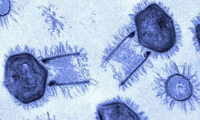
Tupan virus under electron microscope. (Photo: Sci Tech Daily).
Giant viruses are larger than 300 nanometers and can survive waves for millennia. Compared to them, the rhinovirus that causes colds is only about 30 nanometers in size. "The giant virus is massive and extremely complex," said lead author Kristin Parent, associate professor of biochemistry and molecular biology at MSU. "The giant viruses recently discovered in Siberia are still contagious after 30,000 years in the permafrost."
The jagged outer shell of the giant virus helps them to endure harsh environments, protecting the internal genome. The shells of all viruses analyzed in this study have 20 faces. They have a unique mechanism to release the genome. The starfish seal is located at one of the tops of the outer shell. During the infection, both the top and the plug open to release the virus's genome. "Giant viruses are difficult to take pictures because of their size and previous studies rely on finding a virus in an infection state among millions of viruses, " Perent explained.
To solve the problem, graduate student Jason Schrad of Parent's group developed a new method to simulate infection stages. Using the University's new Cryo-EM (Cryo-EM) electron microscope combined with a scanning electron microscope, Parent and colleagues expose giant viruses to exposure to harsh chemical and environmental conditions. similar to what they will experience during the infection process. Cryo-EM allows them to study viruses and protein structures at the atomic level and take pictures while they are active.
The results showed that three environmental conditions successfully promoted the virus to open the peak for infection, namely low pH, high temperature and high salt content. Moreover, each condition triggers a different stage of infection. With the new model, the team discovered that the starfish seal on the top slowly opened while attaching to the shell instead of releasing all the genetic material at once.
With the ability to replicate different stages of infection, Parent and his colleagues studied the viral proteins released in the first stage. Proteins play a part in many biological processes that viruses need to rely on to infect and control the cell machinery that makes copies of them. The team identified important proteins secreted at the beginning of the infection process, helping the virus to complete its invasion of cells.
The fact that many giant viruses respond in the same way in a test tube makes researchers believe that they share similar characteristics and proteins. Unlike corona virus, whether or not the giant virus can infect humans is still a controversial evolution among virologists.
- The newly discovered giant virus can change our definition of a virus
- Science has discovered giant viruses with the ability to "steal" other species' characteristics
- Discovered a giant virus that 'eats alive' bacteria
- Taking advantage of children to infect the virus
- Virus detection makes people silly
- Artificial viruses carry gene molecules and pharmaceuticals into tumors
- Humans have mistakenly thought that the virus is a small creature
- Breakthrough kill metastatic cancer cells
- Smallpox virus can kill cancer cells
- Protein detection inhibits the ability of HIV to infect
- The virus is a living creature and it shares its ancestors with modern cells
- Discover 18 strains of virus originating from mice
- Science makes a virus that kills cancer cells, a major turning point for mankind?
- 14 types of animal infections
 Why do potatoes have eyes?
 'Tragedy' the world's largest carnivorous life: Death becomes ... public toilet
 Tomatoes were once considered 'poisonous' for 200 years
 Are there insects in the figs we eat?