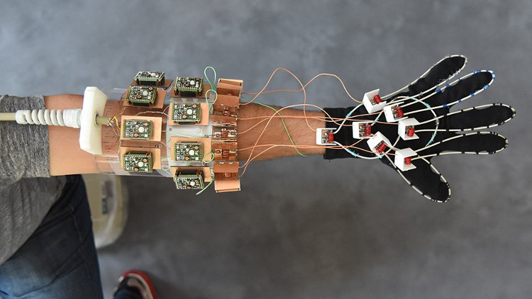
Magnetic Resonance Gloves: A development for hand surgery
Scientists at the New York University School of Medicine are working on making a part of a magnetic resonance imaging (MRI) device designed as a glove to help diagnose future diseases. and allows the construction of flexible atlas images for hand anatomy , which helps guide surgery with more realistic position images or supports the design of better prosthetics .
Limitations of current MRI techniques
Since its first appearance in the 1970s, MRI has helped physicians better observe the tissues inside the body, thus diagnosing millions of cases annually, from tumors in brain to internal hemorrhage or ligament tear. However, this technique has basic limitations.
MRI works by covering tissues in a magnetic field produced when any hydrogen atom aligns to create an average magnetic force in one direction at each slice of tissue. These small "magnets" can then be removed from the balance by electromagnetic waves (radio waves). Whenever this happens, they will rotate (like a spin-off) and also emit a radio signal, allowing the location and visualization to be detected. Thus, the basic principle of MRI is the transmitter. The radio frequency converts radio waves into detectable currents. However, this means that the captured radio waves will produce very few currents inside the signal receiver coil, which will then generate their own magnetic fields and prevent the surrounding coils from capturing clear signals. .
Over the past 30 years, many attempts have been made to manage interactions between broadcast rolls side by side. The result is the introduction of state-of-the-art MRI scanning devices in which signals receive extremely carefully to remove the magnetic field of the surrounding rolls. When the best arrangement is established, the signals will no longer move against each other, which limits MRI's ability to capture complex or moving joints.

MRI signals are generated by hydrogen atoms and therefore this technique is superior in photographing water-rich soft tissue structures.
The problem has now been solved
Since all current MRI scanning devices measure signal generation on detectors, such rolls are always designed in the form of low-impedance structures to allow the current to be transmitted. easily. The new breakthrough of this study is that scientists have designed a structure with high impedance to block the current, and then measure the voltage of the current set in the coil.
When there is no electric current generated by the MRI signal, the new signal-receiving coils will not create a magnetic field that affects the surrounding signal-receiving coils and therefore no longer need hard structures. The researchers found that their system with new signal windings was sewn into a cloth glove, producing sharp images of free mobile muscles, tendons and ligaments in the hands while playing. piano and catch things.
MRI signals are generated by hydrogen atoms and therefore this technique is superior in photographing water-rich soft tissue structures. For that reason, MRI is the ideal technique to photograph tissues, nerves, cartilage . which are difficult to study using other non-invasive methods. However, tendons and ligaments are made of solid proteins instead of liquids, so it is still difficult to see individually because both organizations will appear as black bands running along the bone.
This new study found that, in displaying images of fingers as they bend, the signaling rolls reveal how these black bands move with the bone, which can help classify. There are differences between different types of injuries.
Scientists hope that the results of this study will open a new era in the design of MRI scanners, including the type of bandage around the knee that is injured or the convenient head caps for research. Brain development in newborns.
- Application of elastic magnetic fluids for nuclear magnetic resonance imaging
- The pair of gloves will be the savior of Parkinson's patients
- Magnetic resonance technology replaces cardiac diagnostic techniques
- Wearing a heart aid device can still produce magnetic resonance imaging
- Magnetic resonance imaging tester in milk
- Add fingers for hands
- The new SRS microscope uses MRI technology
- Early magnetic resonance imaging will help save stroke patients
- Magnetic resonance device reaches 90 nm resolution
- Video: Gloves help astronauts as strong as Ironman
- Guess intent through magnetic resonance imaging technique
- Gecko hand glove
 Green tea cleans teeth better than mouthwash?
 Death kiss: This is why you should not let anyone kiss your baby's lips
 What is salmonellosis?
 Caution should be exercised when using aloe vera through eating and drinking