Anatomy of living body in virtual space
A new technology that allows doctors to see clearly a part of your body . This technology helps doctors study diseases, test treatments without having to dissect a living organism . Scientists have designed a computer program that converts medical illustrations into a comprehensive picture of the patient's body, both internal and external.
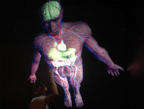
The image is called CAVEman (because it is reproduced in a dedicated room called CAVE), which will not only provide a three-dimensional image of the body and its parts, but it will also show the activity. of those parts, such as the contraction of the heart.
This technology provides an opportunity for physicians and medical students to study diseases, and to test treatments on a ' live ' system without having to dissect a body. living.
Professor Christoph Sensen of biochemistry and molecular biology, and director of the Center for Excellence Center for Visual Genomics of University of Calgary, Alberta (Canada), said: 'We can replicate. a whole new frame is exactly what you see when you treat a patient. '
The system has two main parts : a computer that reproduces the full picture of the body and a virtual environment.

This CAVEman model is reproduced in a dedicated room, called CAVE. In it, equipped with two main parts: a computer that creates a comprehensive picture of the body and a virtual environment (Photo: Masumi Yajima)
This model is based on images in anatomy textbooks. Graphic artists use those images to create the vitality of various systems in the human body, including organs, vascular networks and nervous system. This image is used as a general model of the human body.
This model also has the ability to express a specific human body when mixing the anatomical images of that person. For example, a doctor may combine a patient's CT scan or MRI of the heart or kidney with this pattern, resulting in the patient's organs being visible in a virtual body.
The observation took place in the VAVE dedicated room with an area of about 100 m 2 . Vivid images will be shown on 3 of 4 walls, and also on the floor. Observers are wearing a special type of glass that allows each eye to see each image at the same time. This creates visual fooling of size.
A sensor on the glass incorporates a monitor on the computer to determine the position of the observer as he walks around the virtual body. A joystick allows the viewer to do operations such as turning the virtual pattern or making choices on the menu control panel.
Because this system requires a virtual room for a very expensive cost, Sensen and his team only developed this program on a laptop. However, they only observed patterns in spatial dimensions, but the vibrancy no longer existed.

This model shows the image of a specific person.Observers can enlarge a defined area such as the face and chest area.(Photo: Masumi Yajima)
Anton Koning, research scientist at Erasmus Medical Center (Erasmus Medical Center), in the city of Rotterdam, Netherlands, said: 'I think this is an extraordinary effort to successfully build this model. . Previously, people also used many simple models of human bodies in cinema. '
But this is just the beginning, he stressed: 'The real challenge is when importing data on that virtual body.'
For example, if a researcher wants to reproduce the entire image of a tissue, in which a gene or protein is expressed, the reproduction is quite simple. But reproducing images of many genes at once is extremely complicated. The images then overlap, making observation difficult.
Sensen and her research team have completed the male body model and are finishing the virtual female nodule. After that, they will complete a virtual model of the body of a child as well as other creatures, including some species of mice.
The team also plans to announce their results sometime this summer.
Manh Duc
- See human anatomy, through a unique set of floral images
- Kill patients to save their lives?
- See all day but you probably don't know what this 'hollow' is for
- What would happen if the human body has a structure similar to animals?
- Use VR technology to train surgical skills in place of dead bodies
- Do people have enough living space?
- 8 other applications of virtual reality may appear in the near future
- You will understand the real fear when wearing virtual reality glasses playing horror games when watching this clip
- ISS station astronaut tests HoloLens glasses in space
- Humans will 'move' to space in 2100
- The first transplant patient will become familiar with the new body with virtual reality
- Status of 'virtual body'
 Green tea cleans teeth better than mouthwash?
 Death kiss: This is why you should not let anyone kiss your baby's lips
 What is salmonellosis?
 Caution should be exercised when using aloe vera through eating and drinking