The new biological printing method helps create tissue containing vascular networks
Recently, a team of researchers from the Wyss Institute of Biology at Harvard University introduced a new biological printing method that allows the creation of vascular structures and various cell types. This achievement marks an important step towards creating living tissue with full functionality in the laboratory.
Although 3D printing technology has been used to create human tissue, researchers still have difficulties in regenerating thin tissue layers. Previous attempts to create thicker tissue with a size of one-third of a coin still face many difficulties because the cells inside lack oxygen and nutrients and the cell has no way to discharge. residue. As a result, tissue layers lack air and die.

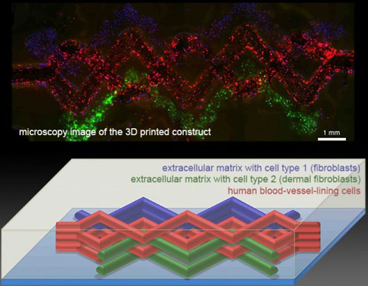
To solve this problem, researchers at the Wyss Institute have used three types of biologically developed inks specially developed with properties borrowed from real living tissue.The first ink uses extracellular matrix , which binds cells together to form tissue. The second ink uses a mixture of extracellular matrix and living cells . The third ink is designed to melt glutinous h. This means that once the team has successfully created the cellular network, it will be cooled, melted and eventually sucked out of the tissue, leaving a network of hollow tubes formed by structure. And this is also the path for blood vessels.
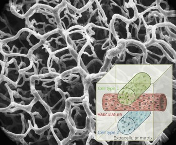
The structure simulates a fundamental property of living tissue, in which the inner cell is maintained by a network of small blood vessels, thin walls, providing oxygen and nutrients and also plays a role of elimination. waste. The team tested simulations and used a model to replicate tissues printed in multiple structures and then create a complex vascular structure from three inks. According to the researchers, this is a structure with very close complexity to human tissue.

3 types of ink corresponding to blue, green and red colors are used to "3D print" the tissue
Professor Don Ingber, director of the Wyss Institute, said: "Tissue regeneration engineers are still waiting for such a method. The ability to shape functional blood vessels in 3D tissues before they are transplanted. only allows the regeneration of thicker tissues, but also increases the ability to link surgery between artificial blood vessels and natural blood vessels to quickly pump blood into transplanted tissues, increasing tissue survival rates. " .
The team's immediate plan for the technology is to focus on creating the closest 3D tissue to live tissue that can be used to test the safety and effectiveness of pharmaceuticals. The Wyss Institute's research has been published in the Advanced Materials issue this month.
- 3D biological printing, pharmaceutical industry revolution
- 3D printed organs bring hope of cheap transplants
- It is possible to create skin layers including blood vessels using 3D biological printers
- Transplantation of biological eye tissue helps blind people see
- Breakthrough technology: Successful 3D printing of human ears with blood vessels and cartilage
- Print human blood vessels with 3D printers
- Research three-dimensional biological printers producing kidneys
- Biological ink breakthrough 'in' human body parts
- Impressed with 3-D nano printers
- Inventing tissue-like materials living with 3D printers
- Stunned modern medicine with newly launched tissue regeneration method
- China successfully implanted 3D blood vessels into monkeys
 Green tea cleans teeth better than mouthwash?
 Death kiss: This is why you should not let anyone kiss your baby's lips
 What is salmonellosis?
 Caution should be exercised when using aloe vera through eating and drinking
 HYBRiD technology makes tissue transparent: Accelerating future disease research
  Breakthrough: Frog regrows amputated leg thanks to special drug
  Scientists have invented a gel that can heal all body and internal wounds
  Found the key to opening the immortal door?
  Use gel from brown algae and soluble polymer to protect donated tissue
  Discover the world's oldest fossil skin in South Africa