China successfully cultivates and implants ears from cartilage cells
Combining 3D printing techniques and cell culture technology, Chinese scientists have succeeded in creating new ears for 5 children with "small ears" - disabilities that affect the shape of ears and listening function.
The researchers said they had collected five children with small ears to feed the new cartilage cell and shaped a new ear for each patient. New ears are cultured in 3D printing in the shape of the healthy ears of patients.
The ear that is raised from the cartilage tissue is then inserted into the affected ear.
The results of this study were published in the January issue of the journal EbioMedicine and were the first to succeed at the same time.
The study wrote: "We have succeeded in shaping, cultivating and reproducing the ear separately for 5 patients". The team suggested that patients should be monitored for 5 years after surgery.
Small ears are a condition in which a child born with one ear develops abnormally or has absolutely no ears - which can weaken hearing. This phenomenon appears at the rate of 1 / 5,000 births, depending on race. Small ears are common in Latin American, Asian, Native American and Andean groups.

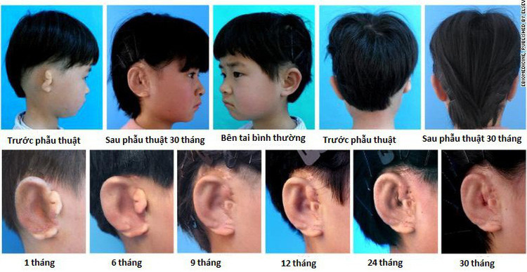
Images of ear implant development and development from cartilage tissue of scientists - (Photo: EBioMedicine).
Common small ear treatment options are plastic prostheses or rib cartilage to create new ears.
Professor Lawrence Bonassar, of biomedical engineering at Cornell University in Ithaca, New York, USA, said: "The ability to shape cartilage for shaping small ears is the goal of the community. science of tissue culture for more than two decades Professor Bonassar did not participate in the research team but was an expert in ear-making from 3D printing techniques ".
"This study shows that tissue culture techniques for ear regeneration and other cartilage tissues will soon become popular in clinics," he said.
The method taken in the newly published study of Chinese scientists is not a new idea. It has been used in mice for many years, since 1997. What's new is that for the first time scientists have applied this method to five patients and followed it for a long time. Five children participated in the program: a six-year-old girl, a 9-year-old girl, an 8-year-old girl, a 7-year-old boy and a 7-year-old girl. All have small ears - the other side is healthy.
Scientists use CT scanners and 3D printers to create a biodegradable biological mold in the shape of a healthy ear. Later, they took cartilage from a small eared ear and kept it in the mold for 3 months to shape the complete ear.
Then they implanted a new ear for the patient and followed it regularly after the transplant. The longest followed patient was 2.5 years.
Of the 5 cases, 4 schools showed very clear cartilage formation after 6 months of ear implant and 3 patients with new ears were the same as healthy ears in size, angle and shape.
Two cases of slight distortion after surgery for ear transplantation.
- Successfully nourish human ears on mice
- Reconstructing ears from patient cells
- Breeding human cartilage in the laboratory
- Chinese doctor implants the ear in the patient's hand
- Japan created a breakthrough in cell technology
- See artificial ear from human ribs
- Video: Tell you how to test your ears yourself
- First implant air graft from stem cells
- Why does salamander grow back almost completely but lizards do not?
- Amazing things from ears
- Successful implantation of trachea develops from stem cells
- How to handle when insects get into their ears
 Green tea cleans teeth better than mouthwash?
 Death kiss: This is why you should not let anyone kiss your baby's lips
 What is salmonellosis?
 Caution should be exercised when using aloe vera through eating and drinking