The method of seeing through organs
Albert Einstein Medical School researchers, Yeshiva University have successfully developed the first fluorescent protein to penetrate the internal organs of living animals without using scalpels or scanning devices. capital with side effects or risk of increased exposure to radiation.
This new body scan method is a big step forward for medical anatomy. For example, this method helps doctors to not 'invade' the patient's body but still check the progress of the tumor, to assess the effectiveness of anti-treatment therapies. cancer.
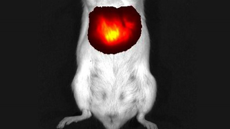
Liver cells in mice experimented with light when iRFP fluorescent protein was present. Photo: Albert Einstein Medical School.
In contrast to other systemic scans, fluorescent protein scans are not associated with the risk of radiation exposure and do not require the use of agents that enhance the contrast of images.
Over the past 20 years, scientists have used a variety of colored fluorescent proteins, jellyfish and coral extracts to lighten the same molecular cells of living cells within living animals. However, the use of fluorescence microscopy methods to project live organism faces great challenges.
The reason is that hemoglobin in animal blood absorbs the wavelengths of green, blue, red and other wavelengths that are used to stimulate conventional fluorescent proteins. Hemoglobin also absorbs any wavelength emitted from proteins when they glow.
To overcome this obstacle, Vladislav Verkhusha, Ph.D., a professor of anatomy and structural biology at Einstein and Dr. Grigory Filonov, author of this paper, designed a fluorescent protein extracted from the substance. phytochrome light receiver. The fluorescent protein based on the new phytochrome, called iRFP, both absorbs and emits light to the near-infrared rays of the spectral line.
In a specific experiment, scientists introduced fluorescent protein into a rat liver - a liver that is particularly difficult to capture because of the relatively high blood volume in the organ. Adenovirus virus molecules containing the gene of iRFP are injected into mice. When these viruses and genes move into liver cells, liver cells produce iRFP protein.
Experimental mice were then exposed to light near the infrared ray and thus could penetrate the liver with fluorescent light emitted. Fluorescence of the liver The experimental mice were first detected on the second day after injecting the iRFP-containing gene. This fluorescence is brightest on the 5th day.
Later experiments demonstrated that the iFFP fluorescent protein was harmless to the body.
' Our research shows that iRFP is much more effective than other fluorescent proteins that are advertised to help with liver scans of living animals ,' said postdoctoral fellow Dr. Grigory Filonov in the laboratory, owner of the paper. Nature Biotechnology magazine said. 'iRFP not only produces much brighter light, with a higher contrast intensity than other proteins but also very stable for a long time. We believe it will be widely used for body scans'.
Dr. Filonov also noted that fluorescent protein scans do not have the potential to be radioactive, often accompanied by computerized tomography (CT) and conventional beam scans. Moreover, unlike magnetic resonance imaging (MRI), it is sometimes necessary to inject a contrast agent into the body, to make the organ look clearer; iRFP screening method does not need to use any additional contrast agent, because of the strong contrast.
The results are published on the electronic version of the journal Nature Biotechnology last July 17.
- India developed a new method of creating organs
- In order to nourish the five organs, it is necessary to
- Eating organs easily accumulates toxins but following these 3 ways will not be toxic
- How to eat organs from animals so that they do not have to be hospitalized?
- 3D printed organs bring hope of cheap transplants
- What kind of organs should be eaten
- Do not worry when the body has an extra spleen
- Detecting the method of embalming the remains of the Egyptian organs
- 7 human organs can be removed and still survive
- Taking organs of pigs to transplant for future people
- Simple test with wrists indicates you can be a model, or sumo fighter
- 6 dangers you face when eating animal organs
 Green tea cleans teeth better than mouthwash?
 Female is XX, male is XY, but why not have YY chromosome?
 Death kiss: This is why you should not let anyone kiss your baby's lips
 What is salmonellosis?